
Human Major Histocompatibility Complex Class II DQ Beta 1 (MHCDQb1) ELISA Kit

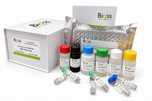
Due to the possibility of mismatching between antigens from other origin and antibodies used in our kits (e.g., antibody targets conformational epitope rather than linear epitope), some native or recombinant proteins from other manufacturers may not be recognized by our products.
Principle of the Assay
The test principle applied in this kit is Sandwich enzyme immunoassay. The microtiter plate provided in this kit has been pre-coated with an antibody specific to Human MHCDQb1. Standards or samples are added to the appropriate microtiter plate wells then with a biotin-conjugated antibody specific to Human MHCDQb1. Next, Avidin conjugated to Horseradish Peroxidase (HRP) is added to each microplate well and incubated. After TMB substrate solution is added, only those wells that contain Human MHCDQb1, biotin-conjugated antibody and enzyme-conjugated Avidin will exhibit a change in color. The enzyme-substrate reaction is terminated by the addition of sulphuric acid solution and the color change is measured spectrophotometrically at a wavelength of 450nm ± 10nm. The concentration of Human MHCDQb1 in the samples is then determined by comparing the OD of the samples to the standard curve.
For use with: Tissue homogenates, cell lysates, and other biological fluids.
For Research Use Only. Not for use in diagnostic procedures.
Target Information
Binds peptides derived from antigens that access the endocytic route of antigen presenting cells (APC) and presents them on the cell surface for recognition by the CD4 T-cells. The peptide binding cleft accommodates peptides of 10-30 residues. The peptides presented by MHC class II molecules are generated mostly by degradation of proteins that access the endocytic route, where they are processed by lysosomal proteases and other hydrolases. Exogenous antigens that have been endocytosed by the APC are thus readily available for presentation via MHC II molecules, and for this reason this antigen presentation pathway is usually referred to as exogenous. As membrane proteins on their way to degradation in lysosomes as part of their normal turn-over are also contained in the endosomal/lysosomal compartments, exogenous antigens must compete with those derived from endogenous components. Autophagy is also a source of endogenous peptides, autophagosomes constitutively fuse with MHC class II loading compartments. In addition to APCs, other cells of the gastrointestinal tract, such as epithelial cells, express MHC class II molecules and CD74 and act as APCs, which is an unusual trait of the GI tract. To produce a MHC class II molecule that presents an antigen, three MHC class II molecules (heterodimers of an alpha and a beta chain) associate with a CD74 trimer in the ER to form a heterononamer. Soon after the entry of this complex into the endosomal/lysosomal system where antigen processing occurs, CD74 undergoes a sequential degradation by various proteases, including CTSS and CTSL, leaving a small fragment termed CLIP (class-II-associated invariant chain peptide). The removal of CLIP is facilitated by HLA-DM via direct binding to the alpha-beta-CLIP complex so that CLIP is released. HLA-DM stabilizes MHC class II molecules until primary high affinity antigenic peptides are bound. The MHC II molecule bound to a peptide is then transported to the cell membrane surface. In B-cells, the interaction between HLA-DM and MHC class II molecules is regulated by HLA-DO. Primary dendritic cells (DCs) also to express HLA-DO. Lysosomal microenvironment has been implicated in the regulation of antigen loading into MHC II molecules, increased acidification produces increased proteolysis and efficient peptide loading.
| GENE ID | 3119 |
| SWISS PROT | P01920 |
| SYNONYMS | HLA-DQB1; HLADQb1; CELIAC1; HLA-DQB; IDDM1; HLA Class II Histocompatibility Antigen,DQ Beta 1 Chain |
Materials Supplied
| Kit Components | 96 Wells Quantity/Size |
|---|---|
| Pre-Coated Microplate | 48T: 6 strips x 8 wells; 96T: 12 strips x 8 wells |
| Standard (Lyophilized) | 48T: 1 vial; 96T: 2 vials |
| Biotinylated Antibody (100×) | 48T: 60 μL; 96T: 120 μL |
| Streptavidin-HRP (100×) | 48T: 60 μL; 96T: 120 μL |
| Standard/Sample Diluent Buffer | 48T: 10 mL; 96T: 20 mL |
| Biotinylated Antibody Diluent | 48T: 6 mL; 96T: 12 mL |
| HRP Diluent | 48T: 6 mL; 96T: 12 mL |
| Wash Buffer (25×) | 48T: 10 mL; 96T: 20 mL |
| TMB Substrate Solution | 48T: 6 mL; 96T: 10 mL |
| Stop Reagent | 48T: 3 mL; 96T: 6 mL |
| Plate Covers | 48T: 1 piece; 96T: 2 pieces |
Storage
| Storage | *If the kit is opened, Store the whole kit at 4°C. If the kit is not used up in 1 week. Store the Pre-Coated Microplate, Standard ,Biotinylated Antibody and Streptavidin-HRP at -20°C, the rest reagents at 4°C, please used up within 6 months. *If the kit is not opened, store the whole kit: 4°C(short time storage, valid for 6 months); -20°C (long-term storage, valid for 1 year). Avoid repeated freeze-thaw cycles. |
Performance Characteristics
| REPEATABILITY | Intra-assay Precision (Precision within an assay):CV% < 8% Three samples of known concentration were tested twenty times on 1 plate to assess intra-assay precision. Inter-assay Precision (precision between assays):CV% < 10% Three samples of known concentration were tested in forty separate assays to assess inter-assay precision. |
| SENSITIVITY | The minimum detectable dose was 0.116ng/mL. |
| ASSAY RANGE | 0.32–20ng/mL |
| SPECIFICITY | This assay has high sensitivity and excellent specificity for detection of Human MHCDQb1. No significant cross-reactivity or interference between Human MHCDQb1 and analogues was observed. |
Frequently asked questions
What is Human Major Histocompatibility Complex Class II DQ Beta 1 (MHCDQb1) ELISA Kit (BSKH73412) used for?
Human Major Histocompatibility Complex Class II DQ Beta 1 (MHCDQb1) ELISA Kit is used for Sandwich Elisa.
Which species does Human Major Histocompatibility Complex Class II DQ Beta 1 (MHCDQb1) ELISA Kit (BSKH73412) detect?
BSKH73412 is designed for Human samples.